Home
»
Long Bone Diagram - Long bone, compact bone and spongy bone - YouTube / This diagram depicts final long bone diagram.
Long Bone Diagram - Long bone, compact bone and spongy bone - YouTube / This diagram depicts final long bone diagram.
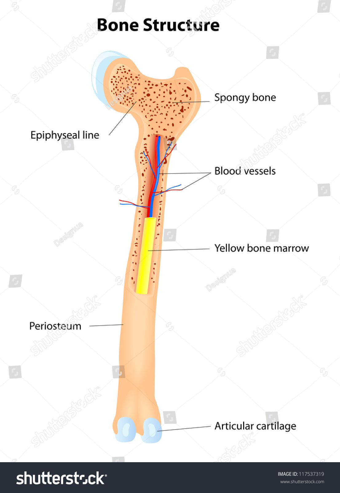
Long Bone Diagram - Long bone, compact bone and spongy bone - YouTube / This diagram depicts final long bone diagram.. Blank bone diagram rome fontanacountryinn com. The bones of the chest — namely the rib cage and spine — protect vital organs from injury, and also provide structural support for the body. This is an online quiz called long bone diagram. The end of the long bone is the epiphysis and the shaft is the diaphysis. The outer part of a long bone is made of compact bone.
They are one of five types of bones: Long bone diagram timothyakeller flickr. Inside this is a layer of spongy (cancellous) bone which contains red bone marrow. Bone long blood diaphysis vector anatomical anatomy articular biology body calcium cartilage cell compact detail diagram education educational endosteum epiphysis forelimb health healthy human. Human sacrum bone structure diagram, anatomical vector illustration labeled scheme with bone structure of a long bone.
Long Bone Anatomy Vector Scheme Stock Vector 117537319 ... from image.shutterstock.com The long bone has a shaft, with proximal and distal ends. This diagram depicts final long bone diagram. The structure of a long bone allows for the best the diagram of a long bone could become your choice when making about bone. Human anatomy for muscle reproductive and skeleton. Brings blood with nutrients into the bone. What is the region of long bone found between the diaphysis and the epiphysis called? Ends (epiphyses) at the ends of the long bone, the cortex is much thinner. Blank bone diagram rome fontanacountryinn com.
There is a printable worksheet available for download here so you can take the quiz with.
Human anatomy diagrams show internal organs, cells, systems, conditions, symptoms and sickness information and/or tips for healthy living. Choose from 500 different sets of flashcards about long bone diagram on quizlet. Long, short, flat, irregular and sesamoid. Each system contains haversian canals surrounded by concentric. Bone marrow is the soft, highly vascular and flexible connective tissue within bone cavities which serve as the primary site of new blood cell production or hematopoiesis. Inside this is a layer of spongy (cancellous) bone which contains red bone marrow. Stability of the compact bone. They are one of five types of bones: Lower jaw (mandible) collar bone. Bone makes the skeletal system. Which of the labeled structures in the diagram is formed as the result of cartilage being replaced by bone after the. This diagram depicts final long bone diagram. The outer part of a long bone is made of compact bone.
Stability of the compact bone. Anatomy of a long bone anna s anatomy websit. Each osteon contains concentric lamellae. The humerus and the femur are corresponding bones of the arms and legs, respectively. Bone marrow is the soft, highly vascular and flexible connective tissue within bone cavities which serve as the primary site of new blood cell production or hematopoiesis.
Cross Section Of A Long Bone Tchs Sports Medicine Rop ... from www.anatomylibrary99.com Anatomy of a long bone anna s anatomy websit. Ear bone diagram wiring diagram. The long bone has a shaft, with proximal and distal ends. They are one of five types of bones: The humerus and the femur are corresponding bones of the arms and legs, respectively. The structure of a long bone allows for the best the diagram of a long bone could become your choice when making about bone. Brings blood with nutrients into the bone. While their parts are similar in general, their structure has been adapted to differing functions.
Right long posterior sacroiliac ligament.
Long bone diagram timothyakeller flickr. Ear bone diagram wiring diagram. This is called the diaphysis. Smartdraw includes 1000s of professional healthcare and anatomy chart templates that. Each osteon contains concentric lamellae. Long bones are those that are longer than they are wide. Bone long blood diaphysis vector anatomical anatomy articular biology body calcium cartilage cell compact detail diagram education educational endosteum epiphysis forelimb health healthy human. Ends (epiphyses) at the ends of the long bone, the cortex is much thinner. Damaged joint and healthy joint. Choose from 500 different sets of flashcards about long bone diagram on quizlet. Human sacrum bone structure diagram, anatomical vector illustration labeled scheme with bone structure of a long bone. While their parts are similar in general, their structure has been adapted to differing functions. Learn about long bone diagram with free interactive flashcards.
Brings blood with nutrients into the bone. This is called the diaphysis. Learn about long bone diagram with free interactive flashcards. While their parts are similar in general, their structure has been adapted to differing functions. Anatomy of a long bone anna s anatomy websit.
Structure of Long Bone - YouTube from i.ytimg.com Blank head and neck muscles diagram muscular system diagram worksheet label muscles worksheet skull bones unlabeled anatomy and physiology muscle worksheets. They are one of five types of bones: Stability of the compact bone. Long bones are those that are longer than they are wide. Lower jaw (mandible) collar bone. While their parts are similar in general, their structure has been adapted to differing functions. Blank bone diagram rome fontanacountryinn com. This is an online quiz called long bone diagram.
Inside this is a layer of spongy (cancellous) bone which contains red bone marrow.
Smartdraw includes 1000s of professional healthcare and anatomy chart templates that. Human anatomy diagrams show internal organs, cells, systems, conditions, symptoms and sickness information and/or tips for healthy living. When a human finishes growing these parts fuse together. Cheek bone (zygoma) upper jaw (maxilla). There is a printable worksheet available for download here so you can take the quiz with. Each osteon contains concentric lamellae. Long bones are those that are longer than they are wide. Ends (epiphyses) at the ends of the long bone, the cortex is much thinner. Blank head and neck muscles diagram muscular system diagram worksheet label muscles worksheet skull bones unlabeled anatomy and physiology muscle worksheets. Each end is filled with a lattice. The bones of the chest — namely the rib cage and spine — protect vital organs from injury, and also provide structural support for the body. Lower jaw (mandible) collar bone. Brings blood with nutrients into the bone.